
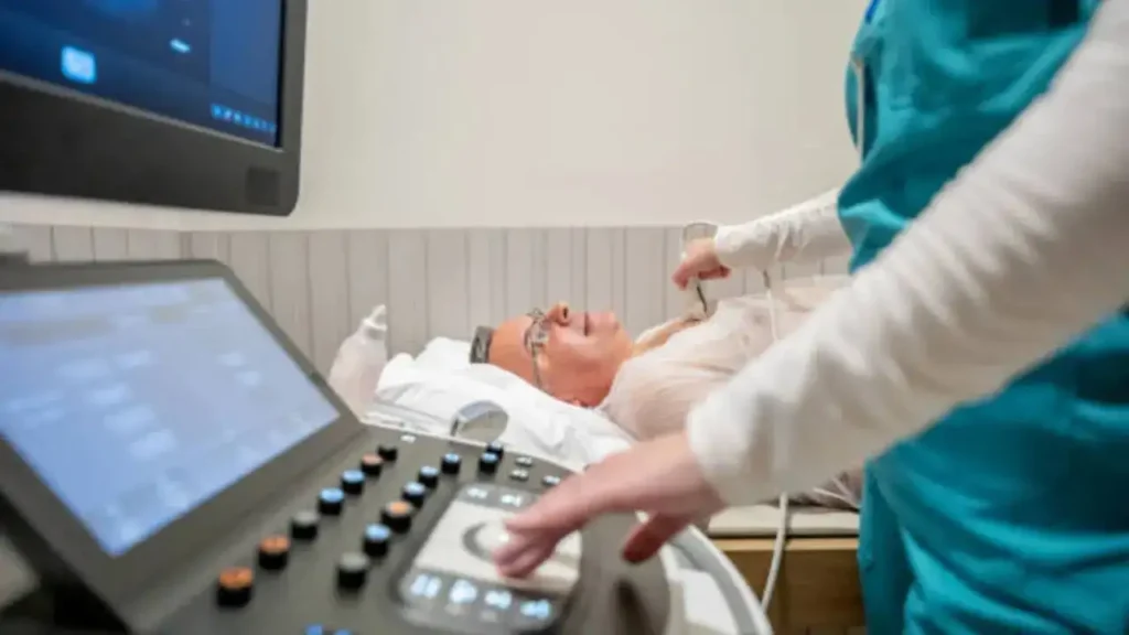
Advances in AI applications in cardiology and echocardiography are rapidly expanding, with the potential to improve patient care. Doctors say AI algorithms help in the detection, classification, diagnosis, and prognostication of cardiac abnormalities and represent a cost-effective tool to address the upsurge in the demand for cardiac imaging.
Echocardiography – also known as the echo test or cardiac ultrasound – is considered the pinnacle of heart-related investigations. Doctors say it provides a rapid, non-invasive, and accurate assessment of heart structure and function, which includes many complex issues as well. However, its widespread availability and low cost profile have pushed it beyond the confines of specialist cardiologists, and it is now a crucial instrument for all clinicians.
But, according to experts, echocardiography also has its own pitfalls. “There are issues like subjective interpretation resulting in poor inter- and intra-observer variation, a longer learning curve, and the need for higher imaging modalities in a complex condition,” Dr M. Sudhakar Rao, Consultant Cardiology, Manipal Hospital, told Times Now.
And so, artificial intelligence-based technology has now emerged to meet these challenges. According to Dr Rao, one of the main advantages of using echocardiography in machine learning models is that its algorithms combine data derived from echocardiography with clinical information and other test results to develop predictive tools. “These are high in accuracy and help enhance diagnosis, risk stratification, and therapeutic strategies,” he said.
AI also uses raw echocardiography images or videos, which can automatically provide structural or functional measurements, along with identifying the state of the disease. “This ability is based on AI’s capacity to automatically analyse features from images that may be too subtle to be detected by the human eye,” said Dr Rao.
What are the applications of AI-powered echocardiography?
A few areas where artificial intelligence in echocardiography can be used include:
- Coronary artery diseases
- Cardiomyopathies
- Cardiac masses
- Valvular heart diseases
- Left ventricular hypertrophy etiology
What is cardiac imaging?
Doctors say cardiac imaging is key to the effective management of patients with coronary artery disease. The use of AI helps us use automated algorithms, which decreases any scope for errors, improves accuracy, and increases specificity and sensitivity.
“Another potential implementation of AI in the field of coronary artery disease could be the differentiation between diseases that commonly present with signs and symptoms similar to an acute coronary syndrome,” said Dr Rao.
AI has also helped in the cases of increased left ventricular wall thickness, where conventional echocardiography may not be sufficient for the etiological diagnoses. AI-based myocardial texture analysis differentiates hypertrophic cardiomyopathy, hypertensive heart disease, athlete’s heart, cardiac amyloidosis, and uremic cardiomyopathy.