Before any cardiac treatment begins, there’s almost always a diagnostic step — and this is often the part patients understand least. A doctor orders an ECG, then maybe an echo, then possibly a stress test, and it’s not always clear why one test wasn’t enough, or what each one is actually looking for. Understanding what these tests reveal makes the whole process far less confusing, and helps you ask better questions during a consultation.
This guide breaks down the most common cardiac diagnostic tests, what each one detects, and when a cardiologist typically orders it. If you’ve been referred for an ECG test in Sarjapur, or you’re trying to understand why multiple tests were recommended, this should clarify the reasoning behind each one.
Electrocardiogram (ECG/EKG): The Starting Point
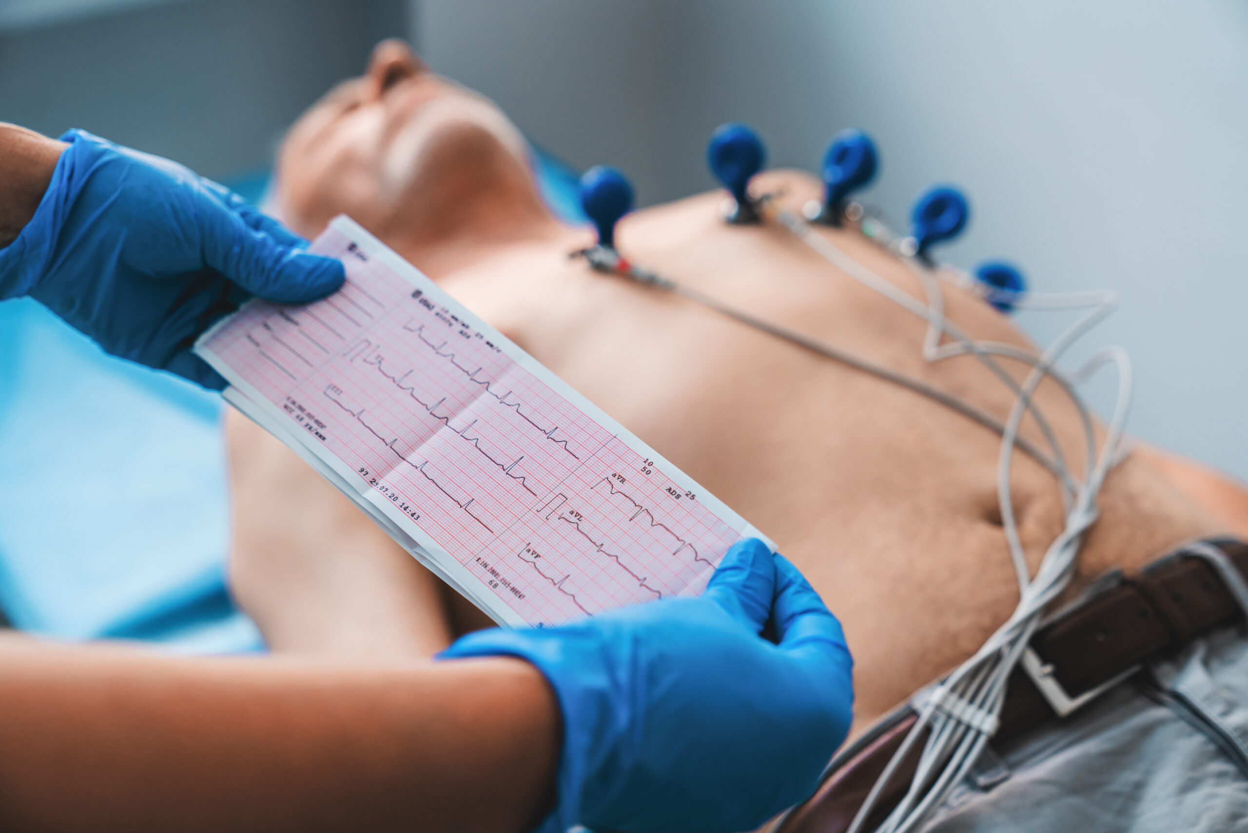
An ECG is usually the very first test in any cardiac evaluation. Electrodes placed on the chest, arms, and legs record the heart’s electrical activity over a short period — typically just a few minutes. It reveals:
- Heart rate and rhythm abnormalities
- Evidence of a previous heart attack
- Signs of an enlarged heart or thickened heart muscle
- Certain electrolyte imbalances that affect heart rhythm
An ECG is quick, painless, and non-invasive, which is why it’s almost always the starting point. However, it only captures a snapshot — a completely normal ECG doesn’t rule out significant coronary artery blockages, since those often don’t produce electrical abnormalities at rest. This is exactly why an ECG is rarely the final word in a proper evaluation.
Echocardiogram (ECHO): Seeing the Heart in Motion
While an ECG captures electrical activity, an echocardiogram uses ultrasound to actually visualize the heart’s structure and movement in real time. For anyone booking an echocardiogram in Sarjapur, this test typically reveals:
- Heart chamber size and wall thickness
- How efficiently the heart is pumping (ejection fraction)
- Valve function — whether valves are narrowing or leaking
- Signs of fluid around the heart
- Early indicators of heart failure
An ECHO is particularly important for anyone with symptoms suggesting heart failure or valve disease, since it directly assesses pumping function and valve behavior in ways an ECG simply cannot.
Treadmill Test (TMT): Assessing the Heart Under Stress
Some blockages only reveal themselves when the heart is working harder. A treadmill test — sometimes called a stress test — monitors heart rate, blood pressure, and ECG readings while you walk or jog on a treadmill at increasing intensity.
A TMT test in Sarjapur is typically recommended when:
- Symptoms like chest discomfort or breathlessness occur specifically during physical activity
- A baseline ECG is normal, but exertional symptoms raise suspicion of blockages
- A cardiologist needs to assess exercise tolerance before clearing someone for a specific activity level
If the test reveals abnormal changes under exertion — ECG changes, chest pain, or an unusual blood pressure response — it often leads to further, more detailed imaging like angiography.
Holter Monitoring: Catching What a Single Test Misses
Many arrhythmias are intermittent — they come and go, sometimes only for a few seconds at a time. A standard ECG, capturing just a few minutes, can easily miss these episodes entirely. Holter monitoring solves this by having the patient wear a small, portable ECG device continuously for 24 to 48 hours (sometimes longer with extended monitors), capturing rhythm data throughout normal daily activity.
This test is typically recommended when:
- A patient reports palpitations, skipped beats, or dizziness that comes and goes
- A standard ECG appears normal despite ongoing symptoms
- A cardiologist needs to confirm the specific type and frequency of an arrhythmia before deciding on treatment
Since arrhythmia treatment varies significantly depending on the specific rhythm abnormality involved, accurately capturing an episode with Holter monitoring is often the deciding factor in choosing the right treatment path.
Cardiac CT: Detailed Structural Imaging Without Invasive Access
A cardiac CT scan uses X-ray imaging combined with computer processing to create detailed cross-sectional images of the heart and its blood vessels. Compared to a standard angiogram, it’s non-invasive — no catheter is required — making it a useful option in specific situations. A cardiac CT scan in Sarjapur is typically used for:
- Assessing coronary artery calcium buildup, an early indicator of plaque
- Visualizing coronary arteries in patients with an intermediate risk profile, before deciding whether invasive angiography is necessary
- Evaluating complex or congenital structural abnormalities
Cardiac CT is particularly valuable as a middle step — more detailed than an ECG or echo, but less invasive than a full angiogram — helping cardiologists decide whether a more invasive procedure is actually warranted.
Cardiac MRI: The Most Detailed Structural Picture
Cardiac MRI uses magnetic fields rather than X-rays to produce highly detailed images of heart structure and tissue composition. It’s particularly valuable for:
- Assessing heart muscle tissue for scarring or damage, including after a heart attack
- Diagnosing certain types of cardiomyopathy (heart muscle disease)
- Evaluating complex congenital heart conditions
- Providing detailed valve and chamber assessment when echo results are inconclusive
Because it takes longer and requires specialized equipment, cardiac MRI is generally reserved for cases where other tests haven’t provided a clear enough picture, rather than being used as a routine first step.
How These Tests Work Together
None of these tests exist in isolation — a good cardiologist selects a combination based on your specific symptoms and risk profile, rather than running every available test by default. A typical diagnostic pathway might look like:
- ECG as the initial baseline assessment
- Echocardiogram if there’s any suspicion of structural or pumping issues
- Treadmill test if symptoms are specifically triggered by exertion
- Holter monitoring if symptoms are intermittent and not captured by a standard ECG
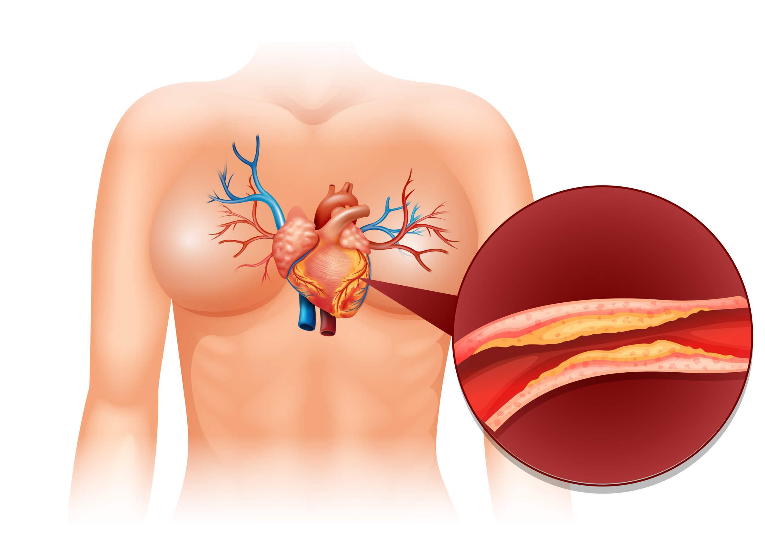
- Cardiac CT or angiography if earlier tests suggest a blockage requiring more detailed visualization
- Cardiac MRI if structural or tissue-level detail beyond echo is needed
This staged approach avoids unnecessary invasive testing while still ensuring nothing significant goes undetected — which is exactly the balance a thorough diagnostic workup should strike.
Why the Right Diagnostic Approach Matters as Much as Treatment
A treatment plan is only as good as the diagnosis behind it. Choosing the wrong test — or stopping after just one when symptoms warrant further investigation — is one of the most common ways early heart disease gets missed. This is why the diagnostic phase deserves just as much attention as deciding between medication, angioplasty, or device implantation once a condition is confirmed.
Dr. M. Sudhakar Rao, a consultant interventional cardiologist practicing at Manipal Hospital, Sarjapur Road, offers comprehensive cardiac evaluation services including ECG, echocardiography, treadmill testing, Holter monitoring, and cardiac CT/MRI — all available in coordination with his interventional expertise, meaning the path from initial diagnostic test to treatment, if needed, happens without unnecessary delays or external referrals.
For more on specific treatments once a diagnosis is confirmed, see the guides on angioplasty, angiography, and device implantation and managing chronic conditions like arrhythmia, valve disease, and hypertension. For more on why facility location and continuity of care matter, see this guide on choosing where to get treated. And if you’re comparing care options by area, related guides are available for Koramangala, HSR Layout, Marathahalli, Bellandur, and Electronic City.
Frequently Asked Questions
Why would I need an echocardiogram in Sarjapur if my ECG was already normal?
An ECG only captures electrical activity, while an echocardiogram shows the heart’s structure and pumping efficiency directly. A normal ECG doesn’t rule out valve problems, pumping issues, or early heart failure, which is why an echo may still be recommended.
Is a TMT test in Sarjapur safe for someone with existing heart problems?
Treadmill tests are generally safe and closely monitored, but your cardiologist will assess whether it’s appropriate based on your specific condition, sometimes opting for alternative stress testing methods if a standard treadmill test carries added risk.
How long do I need to wear a Holter monitor, and does it interfere with daily life?
Standard Holter monitoring typically lasts 24-48 hours, though extended monitoring is sometimes used for less frequent symptoms. The device is small and worn under clothing, allowing most normal daily activities to continue as usual.
What’s the difference between a cardiac CT scan and a cardiac MRI?
A cardiac CT uses X-ray imaging and is faster, often used to assess coronary artery calcium or visualize arteries non-invasively. A cardiac MRI uses magnetic fields and provides more detailed tissue-level information, typically reserved for cases needing a closer look at heart muscle or complex structural issues.
Do I need all of these tests, or just one?
Rarely all at once. A cardiologist typically starts with an ECG and, based on symptoms and findings, adds specific additional tests rather than running a full panel by default, keeping the process efficient and cost-effective.
Can diagnostic imaging alone confirm whether I need angioplasty?
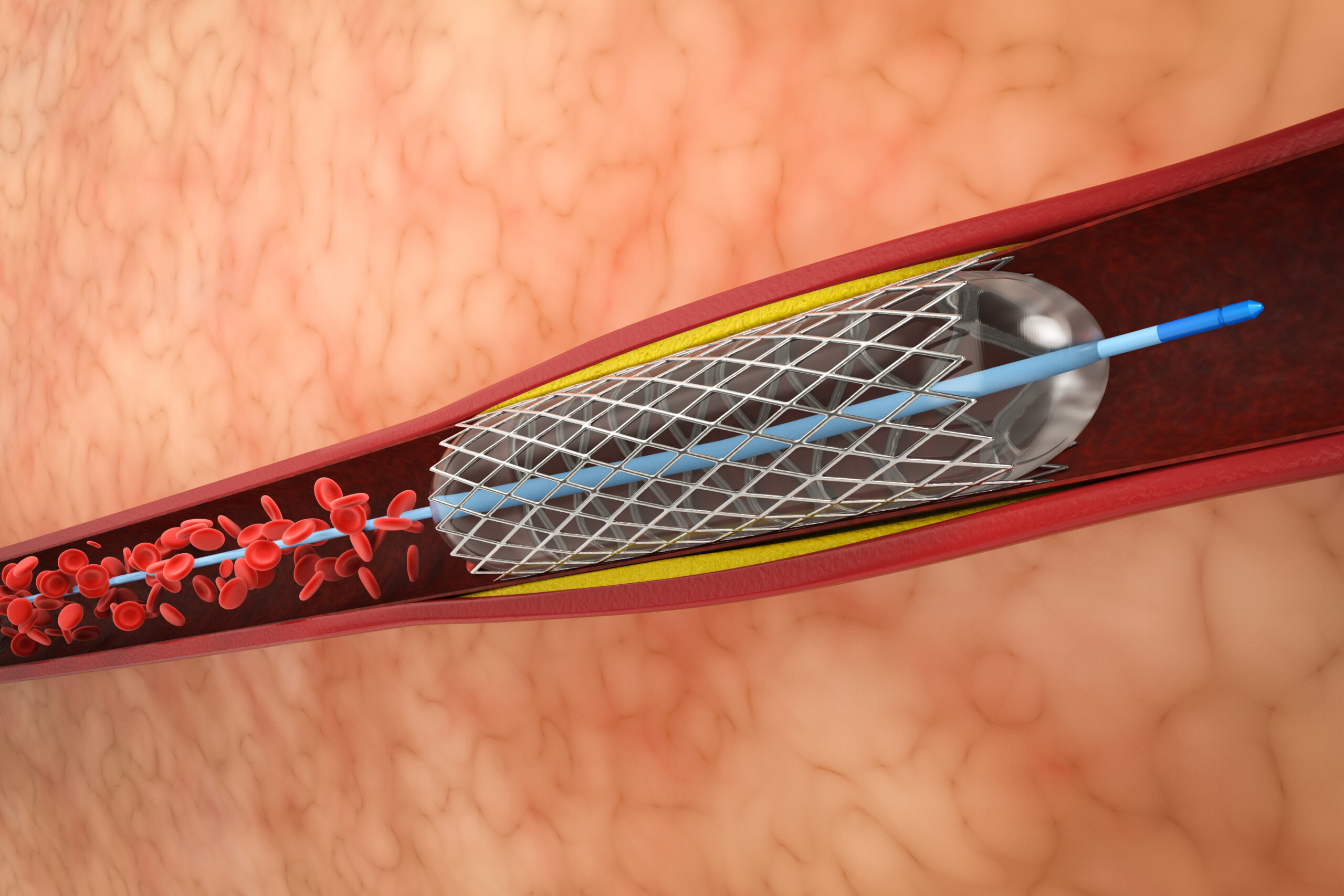
Non-invasive imaging like cardiac CT can suggest the likelihood and location of blockages, but coronary angiography remains the definitive test for confirming severity and deciding whether angioplasty is necessary.
Final Thoughts
Cardiac diagnostics aren’t a single test but a layered process, with each tool answering a different question about heart health. Understanding what an ECG, echocardiogram, treadmill test, Holter monitor, cardiac CT, and cardiac MRI each reveal helps demystify why a cardiologist recommends a specific sequence of tests — and ensures the eventual treatment plan, whatever it turns out to be, is built on an accurate and complete picture of what’s actually happening.